
Darkfield microscopes
Darkfield microscopy illuminates the specimen from an oblique angle so that no direct light enters the objective, only light scattered by the sample itself. This makes unstained, transparent, and low-contrast specimens appear bright against a black background, ideal for viewing algae, plankton, insects, fibers, hairs, yeast, crystals, thin polymers, and some ceramics, and for revealing outlines, edges, and surface detail rather than internal structure. Optika’s darkfield range spans the B-380 and B-510 series, both built around an immersion darkfield condenser with an integrated X-LED illuminator.
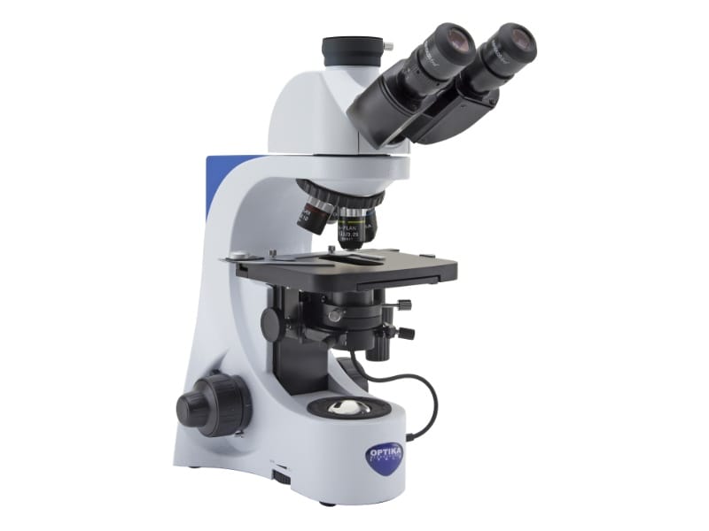
- Type: Darkfield, trinocular
- Magnification: Up to 1000x
- Optics: N-PLAN and W-PLAN objectives
- Illumination: X-LED3 built into darkfield condenser
- Head: Trinocular, 30 degree inclined
€1.874,00 excl. VAT
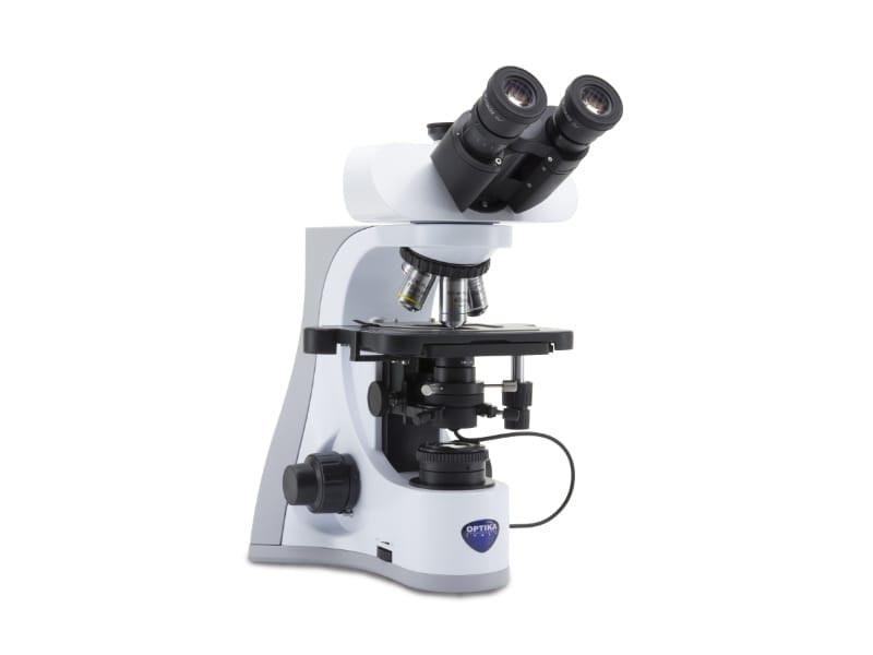
- Type: Darkfield, trinocular
- Magnification: Up to 1000x
- Optics: IOS W-PLAN, infinity corrected
- Illumination: X-LED3, built-in darkfield X-LED
- Head: Trinocular, 30 degree inclined
€2.258,00 excl. VAT
Comparing Optika Darkfield Models
| Model | Series | Optics | Field number | Notable feature |
|---|---|---|---|---|
| B-383DK | B-380 | N-PLAN and W-PLAN 100x oil iris | 20 mm | Built-in X-LED immersion darkfield condenser |
| B-510DK | B-510 | IOS W-PLAN, infinity | 22 mm | Built-in X-LED darkfield condenser with full Kohler brightfield |
How Darkfield Microscopy Works
A darkfield condenser blocks the central light path and directs only oblique light onto the specimen. Light that passes straight through without hitting the sample misses the objective entirely, so the field of view stays dark. Any light that scatters off the specimen, its edges, or internal boundaries is picked up by the objective and appears bright, giving high-contrast images of specimens that would show almost no contrast under standard brightfield illumination, without any staining.
Where Darkfield Is Still Preferred
Phase contrast has replaced darkfield for most general unstained specimen work, since it reveals internal cell structure more accurately and without the halo artifacts darkfield can produce. Darkfield remains the preferred choice in specific situations: fresh, live blood analysis, where its ability to show cell morphology and motion without staining or fixation is well established in hematology; detecting thin, highly motile organisms such as spirochetes, where strong edge-scattering contrast can outperform phase contrast for pure detection; and examining the outlines, edges, and surface features of particulates, fibers, crystals, and low-refractive-index materials, where phase contrast’s halo effect works against clear boundary detection. Darkfield also needs no specialized phase objectives, just a dedicated condenser, which keeps the setup simpler for laboratories focused on these specific applications.
Choosing Between the Series
The B-380 series darkfield model uses N-PLAN objectives with a W-PLAN 100x oil iris objective purpose-built for darkfield oil immersion work, at a mid-range price point. The B-510 series moves to IOS W-PLAN infinity-corrected objectives with a wider 22 mm field of view and full Kohler illumination on the brightfield side, for laboratories that want research-grade optics alongside their darkfield capability. Both models integrate the darkfield condenser’s X-LED illuminator directly, replacing the external 150 W light sources that traditional cardioid darkfield condensers require.
Related Microscope Categories
If darkfield is not the right technique for your sample, see compound brightfield microscopes for stained routine samples, phase contrast microscopes for unstained internal cell structure, polarized light microscopes for birefringent minerals and crystals, upright fluorescence microscopes for immunofluorescence work, or upright metallurgical microscopes for opaque metal and ceramic samples.
For the full upright range overview, see upright microscopes.